Microscopy
Overview
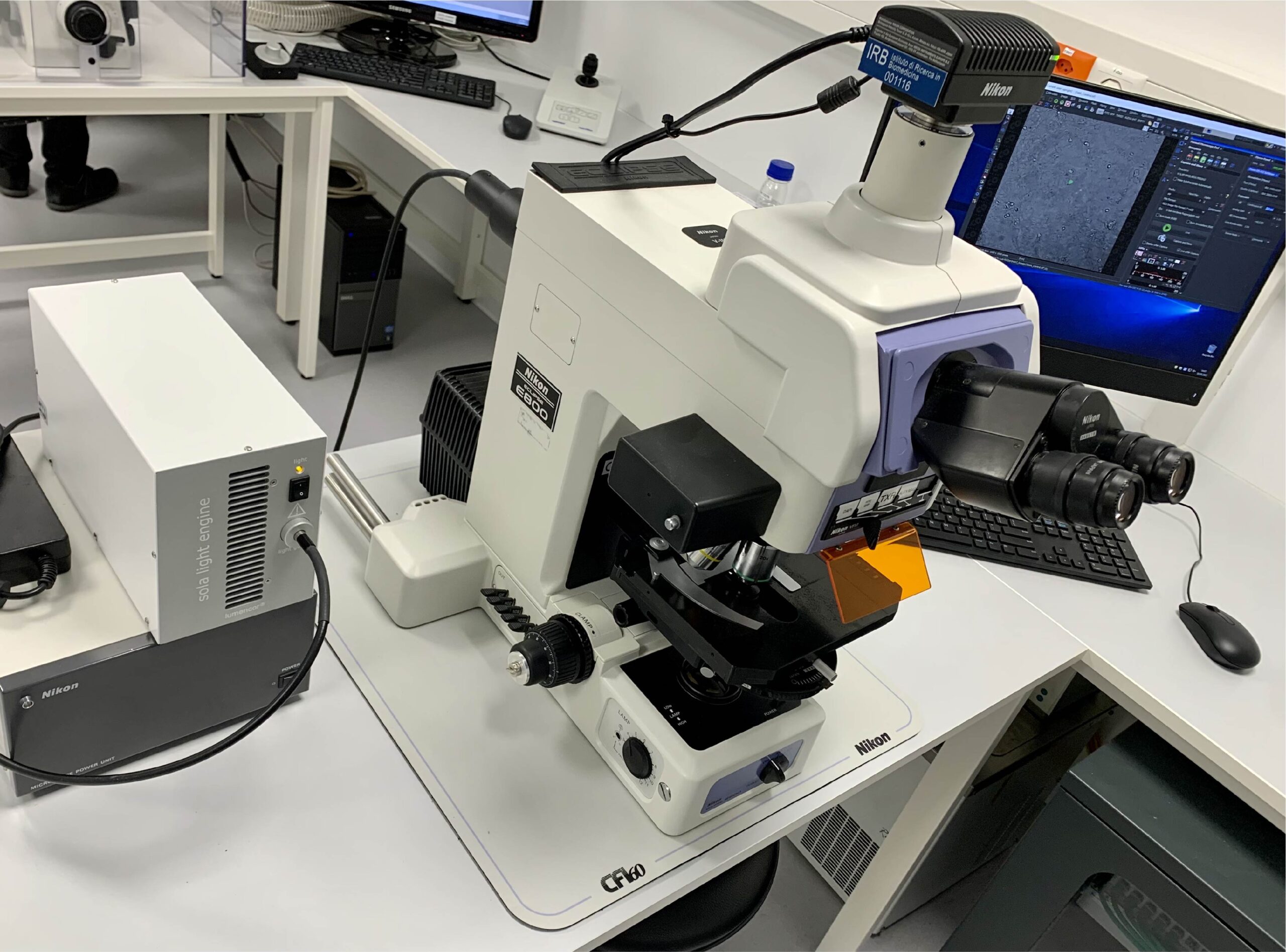
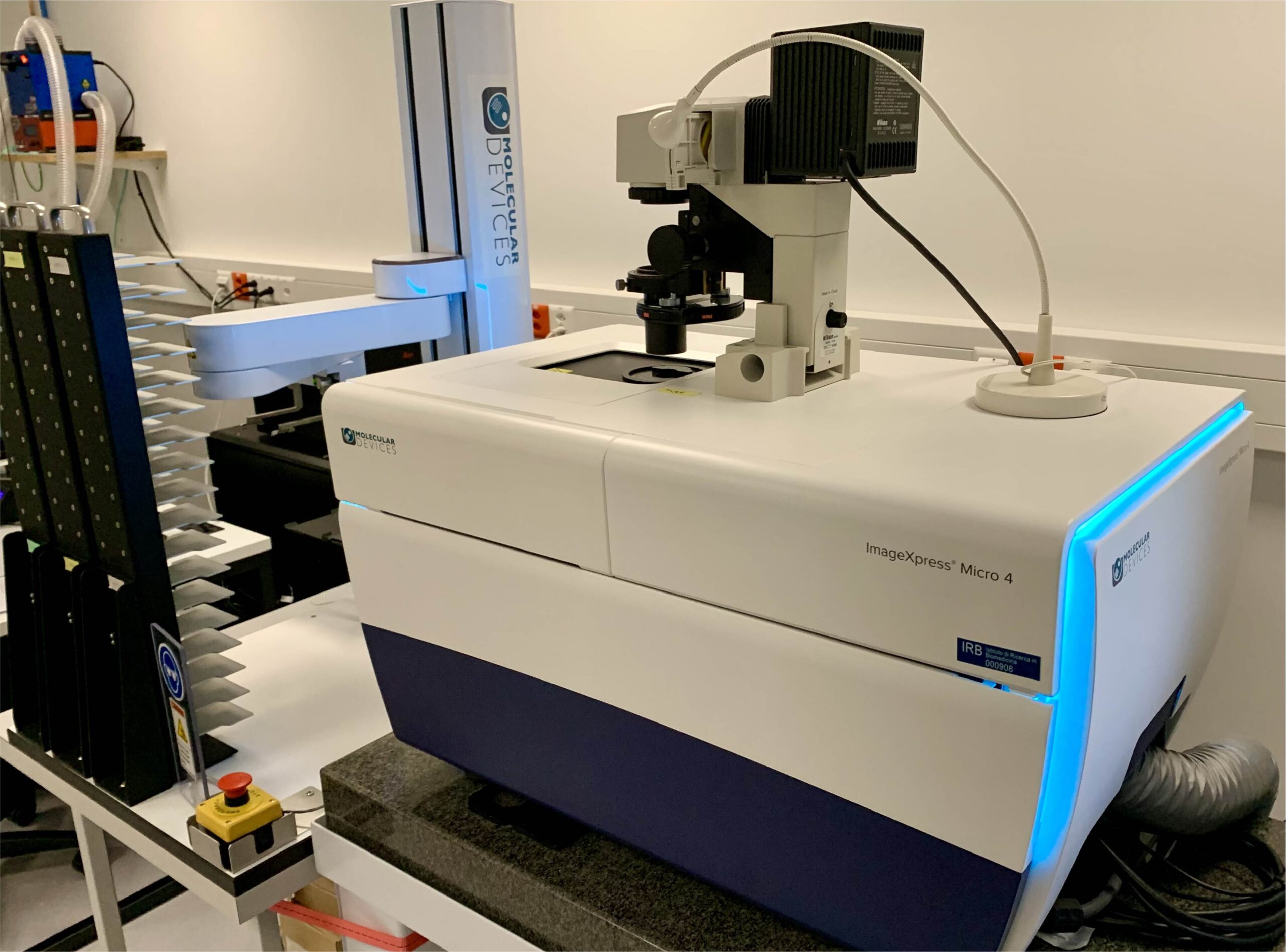
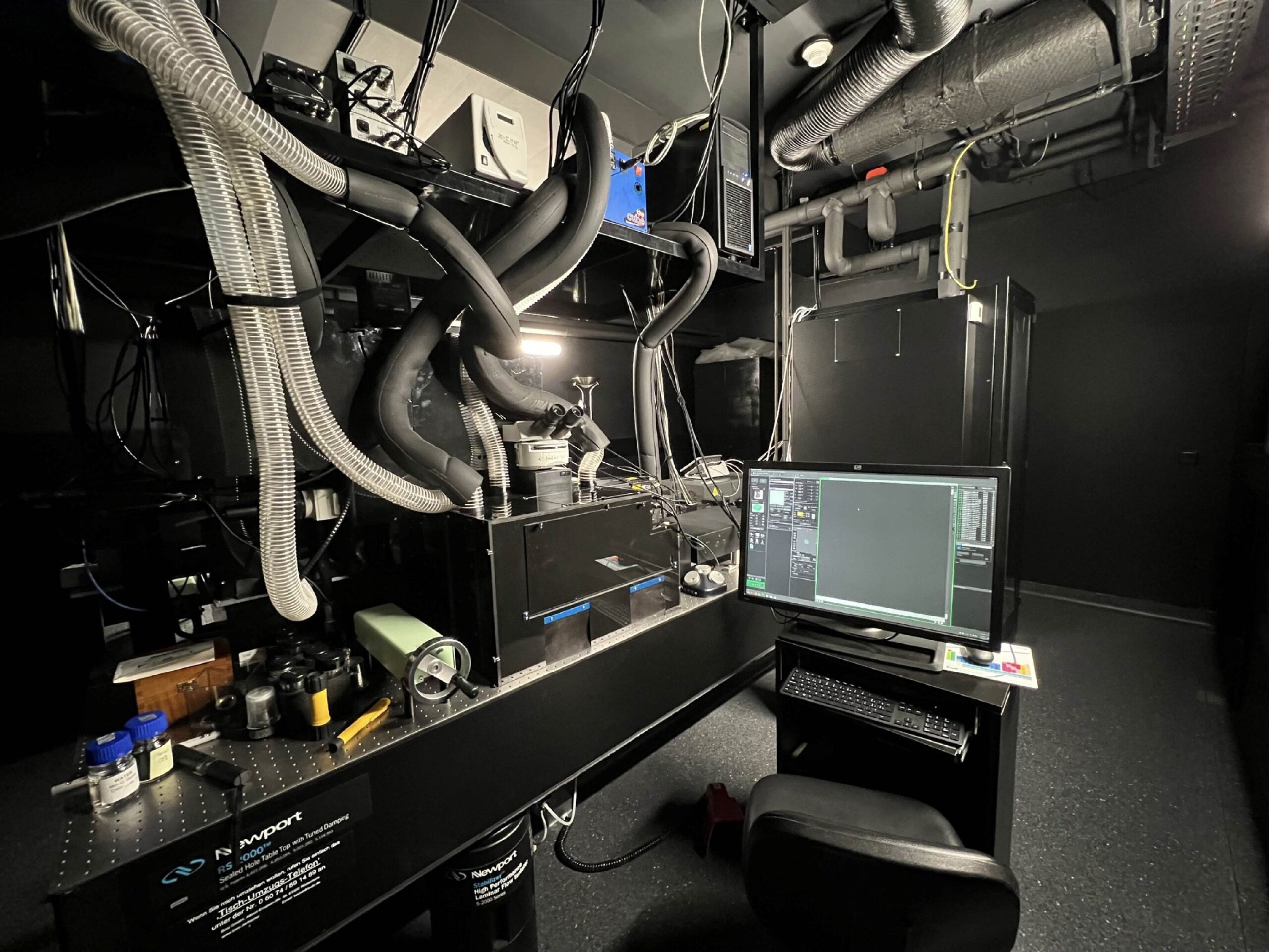
The facility provides state-of-the-art imaging equipment and expertise for the most demanding research projects from subcellular events to cell dynamics in organs. We also offer support and courses for students and researchers.
Equipment
Key technical features
– Imaging lasers 405, 488, 561, 642 nm
– Photomanipulation laser two-photon 920 nm
– Sheet thickness from 1.5 µm to 2.2 µm
– Magnification changer from 12.5x to 50x
– Uniform illumination module
– Destriping module
– Mosaic module
– Sample tracking module
– Python-controllable API for feedback microscopy
Key technical features
– 2 Chameleon Coherent tunable lasers 690-1040 nm
– Coherent OPO 1000-1300 nm
– Double-headed upright and inverted
– 3 EOMs for photomanipulation
– Objectives 4x to 100x
– 5 GaAsP detectors for each side
– Incubator
– Python-controllable API for feedback microscopy
With these systems, we can perform most cell and tissue imaging procedures, such as mosaic reconstructions of tissues and organoids, STED, fast live-cell and organoid imaging, FRAP, photoconversion, FLIM-FRET, and 3D reconstruction on unclarified and clarified tissues. Images with these techniques can also be combined with Electron Microscopy micrographs in Correlative Electron Microscopy (CLEM).


Support
Courses
The facility is also open to collaborations, such as the development of new tools, techniques and analysis software. An example is LysoQuant.